Heart Anatomy

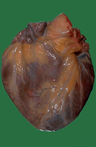
This is the external appearance of a normal heart.
The epicardial surface
is smooth and glistening.
The amount of epicardial fat is usual.
The left
anterior descending coronary artery extends
down from the aortic root to the
apex.

From the moment it begins beating
until the
moment it stops, the human heart works tirelessly.
In an average
lifetime, the heart beats more than
two and a half billion times, without
ever
pausing to rest. Like a pumping machine,
the heart provides the power
needed for life!!

1.Right Coranary
9.Right Atrium
2.Left Anterior Descending 10.Right Ventricle
3.Left
Circumflex 11.Left Atrium
4.Superior Vena Cava 12.Left
Ventricle
5.Inferior Vena Cava 13.Papillary Muscles
6.Aorta 14.Chordae
Tendineae
7.Pulmonary Artery 15.Tricuspid Valve
8.Pulmonary Vein 16.Mitral
Valve
Aortic Valve(not pictured) 17.Pulmonary
Valve

The heart you see drawn on the
average Valentine
is only a rough representation of the actual
structure
of the heart. Your heart is
actually shaped more like an upside-down
pear.
The human heart is primarily a shell. There
are four cavities,
or open spaces,
inside the heart that fill with blood.
Two of these
cavities are called atria.
The other two are called ventricles.
The two
atria form the curved top
of the heart. The ventricles meet at the
bottom
of the heart to form a pointed
base which points toward the left side
of
your chest. The left ventricle contracts
most forcefully, so you can best
feel
your heart pumping on the left side
of your chest.
The left
side of the heart houses one
atrium and one ventricle. The right side
of
the heart houses the others. A wall,
called the septum, separates the right
and
left sides of the heart. A valve
connects each atrium to the
ventricle below it.
The mitral valve connects the left atrium
with the
left ventricle. The tricuspid valve
connects the right atrium with the right
ventricle.
The top of the heart connects to a few
large blood
vessels. The largest of these
is the aorta, or main artery, which
carries nutrient-rich blood away from the heart.
Another important vessel
is the pulmonary artery
which connects the heart with the lungs
as part of
the pulmonary circulation system.
The two largest veins that carry blood
into the heart are the superior vena
cava and the inferior vena cava.
They
are called "vena cava" because they are
the "heart's veins." The
superior is located
near the top of the heart.
The inferior is located
beneath the superior.
The heart's structure makes it an
efficient,
never-ceasing pump. From the moment of
development through the
moment of death,
the heart pumps. The heart, therefore,
has to be strong.
The average heart's
muscle, called cardiac muscle, contracts and
relaxes
about 70 to 80 times per minute
without you ever having to think about
it.
As the cardiac muscle contracts it pushes
blood through the chambers
and into the vessels.
Nerves connected to the heart regulate the
speed
with which the muscle contracts. When you run,
your heart pumps more
quickly. When you
sleep, your heart pumps more slowly.
Considering
how much work it has to do,
the heart is surprisingly small. The
average
adult heart is about the size of
a clenched fist and weighs about 11
ounces (310 grams). Located in the middle
of the chest behind the
breastbone, between
the lungs, the heart rests in a
moistened chamber
called the pericardial cavity which
is surrounded by the ribcage. The
diaphragm,
a tough layer of muscle, lies below.
As a result, the heart is
well protected.
Function of the Heart
Every cell in your body
needs oxygen in order
to live and function. The role
of the heart is to
deliver the
oxygen-rich blood to every cell in the body.
The arteries are
the passageways through which
the blood is delivered. The largest
artery
is the aorta, which branches off the
heart and then divides into many smaller
arteries.
The veins carry the deoxygenated blood
back to the lungs to pick
up more oxygen,
and then back to the heart once
again. Blood flows
continuously through the
circulatory system, and the heart muscle is
the
pump which makes it all possible!
Coronary Arteries
Your heart,
just like all other muscles
in the body, needs its own supply of
oxygen in
order to function properly. Although
its chambers contain blood, the heart
receives no
nourishment from the blood inside the chambers.
The heart gets
its blood supply from the
coronary arteries. The two major
coronary
arteries (the right coronary artery and the
left main coronary
artery) branch off the aorta,
and then divide into many smaller
arteries
that lie in the heart muscle and feed the heart.

For
further information contact:
National Heart Lung, and Blood Institute
(NHLBI)
Information Center
P.O. Box 30105
Bethesda MD
20824-0105
Don't be afraid your life will end,
BE AFRAID it will never
begin!!
Please sign my
guest book and let me know what you think!
>
New Guestbook, Images allowed
 Back Home